Introducing iQ Slice™ & iQ Fan™
The two features harness the advanced proprietary ultrasound chip matrix integrated into the latest Butterfly Ultrasound whole-body handheld ultrasound probe. These unique features are unparalleled in handheld ultrasound technology, utilizing the chip matrix to sequentially activate arrays, thereby generating multiple ultrasound planes from a single probe position.
This innovation reduces the need for continual probe adjustments during organ system scans. It represents not only a technological leap forward that improves scanning procedures for practitioners but also serves as an invaluable educational resource. Learners get a comprehensive view of 3D anatomy by scrolling through sequential ultrasound cross-sections., fostering a deeper understanding of anatomical orientation within ultrasound imagery.
In this paper, we explore the technical intricacies of the iQ Slice™ and iQ Fan™ technology. We examine its integration with common clinical scanning protocols, highlighting its capacity to enhance diagnostic efficiency by providing "more information with less movement". Additionally, we elucidate its role as a potent educational instrument, aiding in the understanding of ultrasound plane cross-sections and their implications for anatomical interpretation.
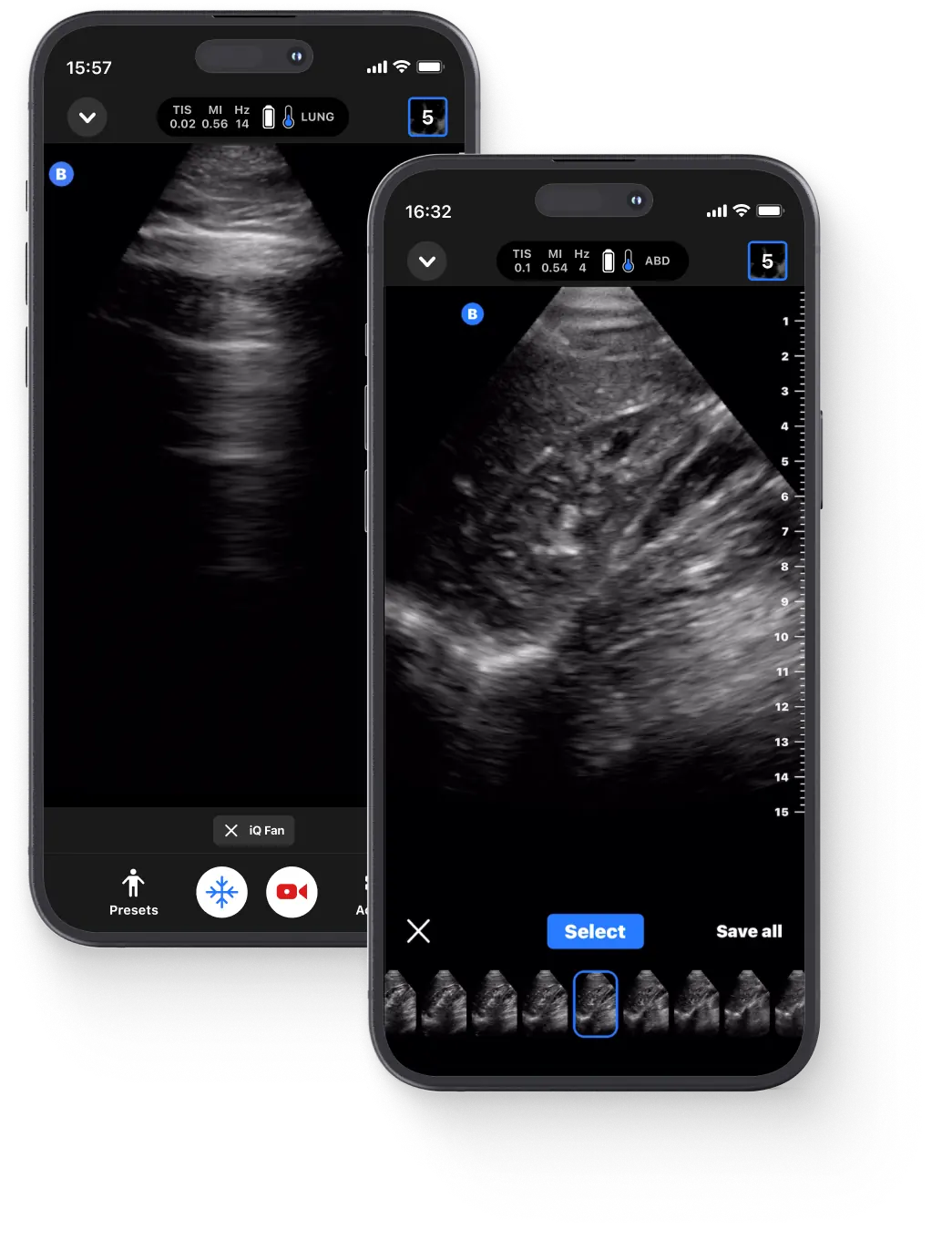
Butterfly Network revolutionized traditional ultrasound transducer technology.
By integrating a singular silicon chip hosting a matrix array of Capacitive Micromachined Ultrasound Transducers (CMUTs) into a handheld ultrasound probe (the Butterfly iQ probe). These CMUTs function by utilizing a conductive membrane and substrate forming a capacitor separated by a vacuum gap. When voltage is applied, it causes the membrane to vibrate, thereby generating ultrasound waves. In turn, reflected sound waves cause the membrane to vibrate as well, which are then captured as electric signals and converted into images.
What sets CMUT technology apart from traditional piezoelectric crystals is its ability to emit and detect soundwaves across a wide range of frequencies, offering a broader bandwidth in a single probe.s. By arranging CMUTs in a matrix array, the Butterfly iQ can emulate different transducer types—linear, curved, and phased —within a single probe, facilitating versatile whole-body imaging capabilities. This array configuration is instrumental in enabling 3D imaging within the ultrasound domain.
The Butterfly iQ's integration of CMUT technology with electronic circuits condenses probe-to-control wiring into a semiconductor chip, significantly reducing complexity and enhancing the probe's portability. This technological advancement, achieved through the Complementary Metal-Oxide Semiconductor (CMOS) process, enables cost-effectiveness and scalability in ultrasound production.
The latest Butterfly ultrasound probe, the iQ3, represents a significant advancement over its predecessor, the iQ+, primarily due to its state- of-the-art P4.3 chip. This new chip delivers faster beamforming, resulting in improved data transfer rates, which in turn leads to enhanced 3D capabilities and superior imaging. The iQ3's rapid processing capabilities translate into a better frame rate, significantly elevating the quality of real-time imaging. With enhanced post-processing capabilities, clinicians can obtain images of unparalleled quality, making the iQ3 a groundbreaking innovation in chip-based ultrasound technology.
Moreover, the exclusive integration of Butterfly Ultrasound's proprietary technology enables the iQ Slice and iQ Fan features. These features leverage CMUT matrix capabilities to sequentially activate arrays, generating multiple ultrasound planes from a single probe position. This allows for a broader understanding of anatomical orientation and facilitates efficient scans across extensive anatomical sections without the need for frequent manual probe manipulation. As illustrated in Figure 1, the iQ Slice feature enables ultrasound scans to encompass a 1.68cm cross-sectional width for each 1.Ocm of depth within the body. This translates into the iQ Slice feature seamlessly scanning an 8.4cm width at a depth of 5cm and an impressive 16.8cm width at a depth of 10cm (Figure 1).

This innovative approach offers learners a comprehensive perspective on anatomical orientation concerning ultrasound cross-sections, enhancing the comprehension of complex anatomical structures and their spatial relationships. Moreover, much like how a high-radiation CT scanner offers cross-sectional views of anatomy that can be selectively examined, the iQ Slice tool extends this capability to a handheld ultrasound device (Figure 2).
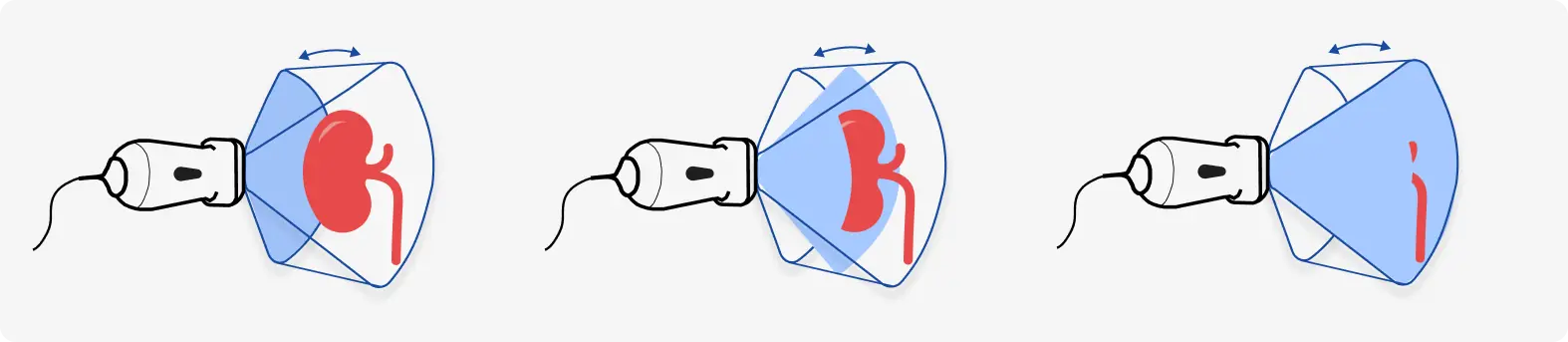
iQ Slice™ and iQ Fan™, both harness the power of Butterfly's unique CMUT matrix, expanding the possibilities of ultrasound imaging.
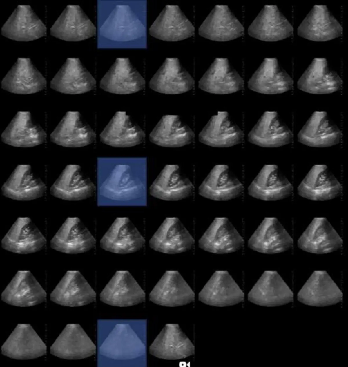
The technology of iQ Fan"' can also be utilized in a unique manner, engaging the iQ Slictechnology in a repeated fashion over a 6-second time interval. This feature has been aptly labeled "iQ Fan it allows the user to obtain repeated "iQ Slice" scans, mimicking the act of fanning back and forth across the organ of interest.
These offerings, iQ Slice and iQ Fan, both harness the power of Butterfly's unique CMUT matrix, expanding the possibilities of ultrasound imaging and providing healthcare practitioners with versatile tools for efficient, detailed, and dynamic examinations. These features showcase the exceptional capabilities of the Butterfly iQ3, differentiating it from conventional ultrasound systems and making advanced imaging accessible through an economical, handheld probe.
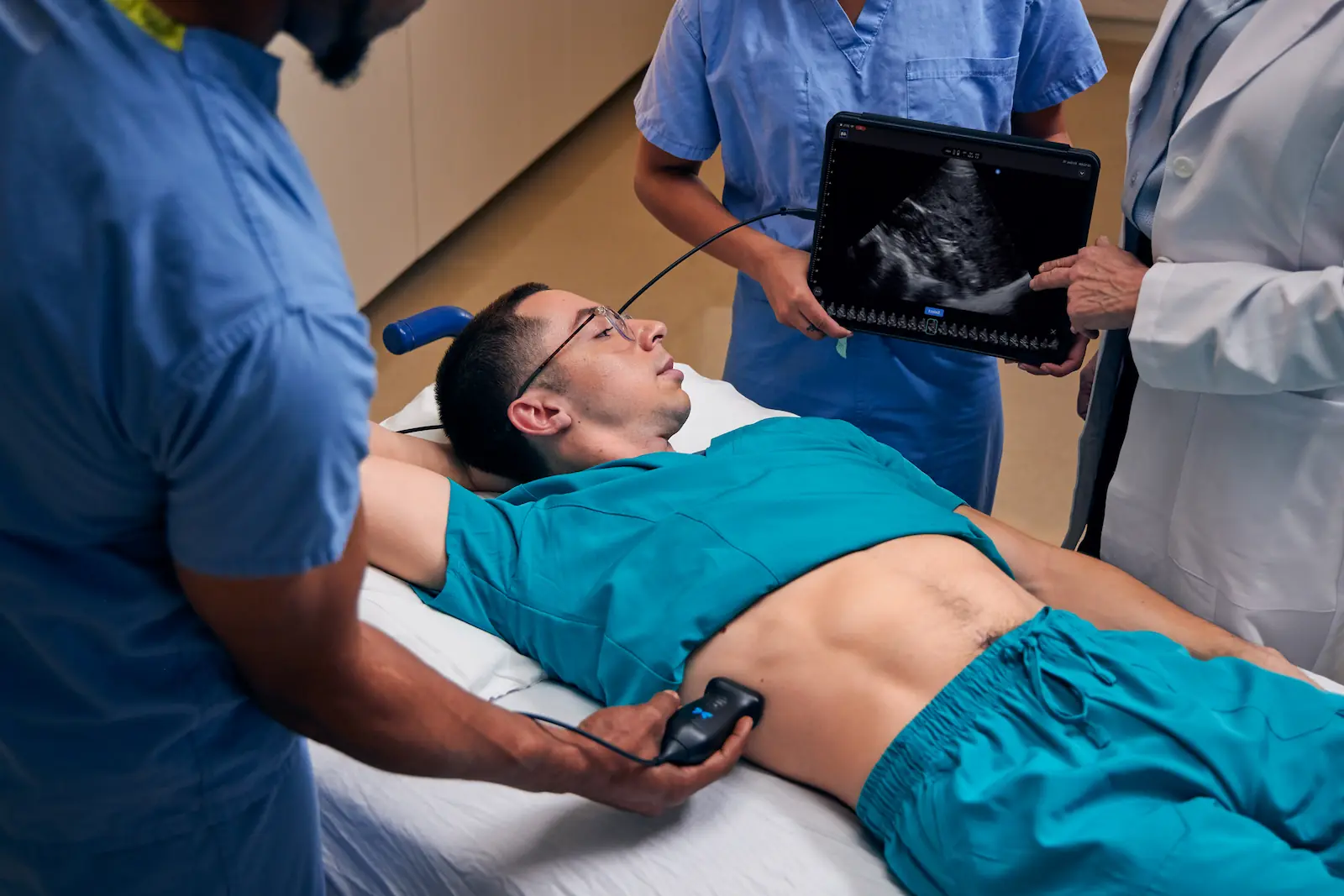
Application of iQ Slice™ into Clinical Scanning Protocols.
Within abdominal ultrasound scans, current protocols require a series of scans in different orientations to comprehensively evaluate various anatomical structures. For instance, in liver assessment, the American Institute of Ultrasound in Medicine (AIUM) Liver Ultrasound Guidelines [1] involve static scans in both sagittal and transverse planes, capturing images at 1-cm increments. This extends to real-time scans covering the diaphragm, hepatic veins, portal veins, bile ducts, fissures, ligaments, lobes, gallbladder, and right kidney, each necessitating separate probe position adjustments.
iQ3 Slice would efficiently streamline the abdominal scanning process. While probe placement in each orientation remains necessary, iQ Slice allows for the sequential acquisition of multiple slices in each orientation within a single scan, reducing the number of manual sweeps and adjustments required.
This has the potential to expedite the scanning process, enabling a comprehensive overview of liver anatomy and pathologies.
The iQ Slice feature not only enhances liver assessments but also presents potential advantages in additional clinical scanning protocols, including gallbladder and biliary tree examinations, renal evaluations, and abdominal aorta screening. Table 1 provides a comprehensive overview of the benefits of iQ Slice Technology in abdominal scanning protocols.
iQ Slice™ allows for the sequential acquisition of multiple slices in each orientation within a single scan, reducing the number of manual sweeps and adjustments required.
The iQ Slice feature potentially enhances the efficiency of these abdominal scans by reducing the number of sweeps and adjustments required while offering a comprehensive assessment of anatomical structures, revolutionizing traditional scanning protocols. This first-of-its-kind feature is provided not by a high- end ultrasound system but rather by the economical whole-body ultrasound probe from Butterfly Network, showcasing the remarkable technology accessible within this portable and cost-effective device.
Benefits of iQ Slice™ Technology in Abdominal Scanning Protocols.
Application of iQ Fan™ for Pulmonary Ultrasound
The application of the iQ Fan™ feature for thoracic ultrasound represents an advancement in ultrasound imaging for the evaluation of lung pathologies.
Traditional pulmonary ultrasound requires dynamic ultrasound plane adjustments to detect the common artifacts used for pathology detection, A-lines and B-lines. These artifacts provide essential diagnostic information but demand meticulous scanning and adjustments by the sonographer. Additionally, the detection and quantification of pleural effusions also require similar dynamic probe adjustments.
The iQ Fan" feature mimics this dynamic technique, empowering iQ3 users by effectively decreasing their scanning burden. By automating the fanning motion, users can focus more on image interpretation and streamline the thoracic ultrasound examination process. This innovation bridges the gap between complex thoracic evaluations and user-friendly, handheld ultrasound technology, enhancing the efficiency and diagnostic capabilities of thoracic ultrasound examinations.
In a clinical study, a board-certified Internal Medicine Resident at Ascension Health stated that iQ Slice made a "significant improvement in their scanning confidence and efficiency", They remarked that the technology allows for "faster capture" by "eliminating the need for precise probe adjustments, thereby enhancing the accuracy of anatomical detail capture."
Improving Point- of-Care Ultrasound (POCUS) education represents a critical aspect of advancing medical training in the field of ultrasound.
A common challenge lies in teaching learners how ultrasound planes insonate organ systems and the precise probe manipulations required to scan them effectively. Traditional methods of ultrasound education often involve lengthy training periods and significant hands-on experience to grasp the nuances of acquiring diagnostic images.
The complex relationship between probe orientation, ultrasound beam angle, and resulting image is a substantial barrier to learning. iQ Slice technology within the Butterfly iQ3 presents a revolutionary solution to this challenge. By enabling learners to visualize multiple ultrasound planes sequentially, iQ Slice provides a comprehensive understanding of how probe manipulation impacts image acquisition. This approach allows learners to grasp the intricacies of organ system examination more efficiently. iQ Slice transforms ultrasound education by offering a dynamic, real-time learning experience, bridging the gap between theoretical knowledge and practical proficiency in POCUS. It empowers learners to develop the necessary skills to navigate the complexities of ultrasound examinations effectively, ultimately enhancing the quality of patient care and diagnostic accuracy.
This innovative technology holds the potential to transform the landscape of ultrasound.
Butterfly Ultrasound's iQ Slice and iQ Fan tools, integrated into the iQ3 handheld ultrasound probe, represent groundbreaking advancements in both medical imaging and education.
These technologies harness the power of CMUT to provide users with a comprehensive view of 3D anatomy, a streamlined diagnostic processes and an enhanced learning experience. iQ Slice™ and its complement, iQ Fan™, showcase the remarkable capabilities of the Butterfly iQ3, making advanced imaging accessible through an economical, handheld probe. This innovative technology holds the potential to transform the landscape of ultrasound, improving patient care and education in the field of medical imaging.
Image 1. Normal Side. Pediatric Lung setting, demonstrates clear pleural line with sliding and z lines (aka comet tails). No indication of B lines; normal appearing lung
Image 2. Abnormal side. Additional air bronchograms further identifying the consolidated lung region. With respiration consolidated lung is partially obscured by B lines. Consolidated lung with dynamic air bronchograms
Image 3. Abnormal Side. On left of image: few B lines indicating presence of fluid in lung, irregularly appearing pleural line with sub pleural consolidation (~ 2 cm in depth). Consolidated lung area looks like liver hence the term, lung hepatization. Echogenic dots are air bronchograms; these collections move with respiration therefore they are called dynamic air bronchograms. (Note: dynamic air bronchograms = pneumonia; static air bronchogram - no movement with respiration = atelectasis)
References:
- (2022), The AIUM Practice Parameter for the Performance of an Ultrasound Examination of the Abdomen and/orRetroperitoneum. J Ultrasound Med, 41: E1-E8.
- (2022), The AIUM Practice Parameter for the Performance of an Ultrasound Examination of the Abdomen and/orRetroperitoneum. J Ultrasound Med, 41: E1-E8.
- (2022), The AIUM Practice Parameter for the Performance of an Ultrasound Examination of the Abdomen and/orRetroperitoneum. J Ultrasound Med, 41: E1-E8.
- (2021), AIUM Practice Parameter for the Performance of Diagnostic and Screening Ultrasound Examinations of theAbdominal Aorta in Adults. J Ultrasound Med, 40: E34-E38.