A normal X-ray, a physical job, and a full-thickness tear
Introduction: The diagnostic gap
An X-ray can rule out fracture, but it cannot image tendon, which is how patients with serious tendon injuries get sent home being told by a physician, "you probably just strained something". Ultrasound images the tendon directly and, pooled against surgical findings across 65 studies, is comparable to MRI in sensitivity and specificity for partial and full-thickness rotator cuff tears.1
Presentation
50-year-old male, construction worker, fell backward off a ladder onto an outstretched arm. Urgent care radiograph read normal; discharged with a sling, analgesia, and a presumed strain. Days later he had not improved, could not raise his arm to carry lumber or shingles, and was not working.
Examination
Provocative testing was positive but non-discriminating. It implicated the cuff without characterizing the lesion.2
Sonographic findings
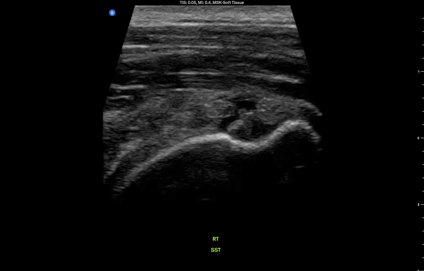
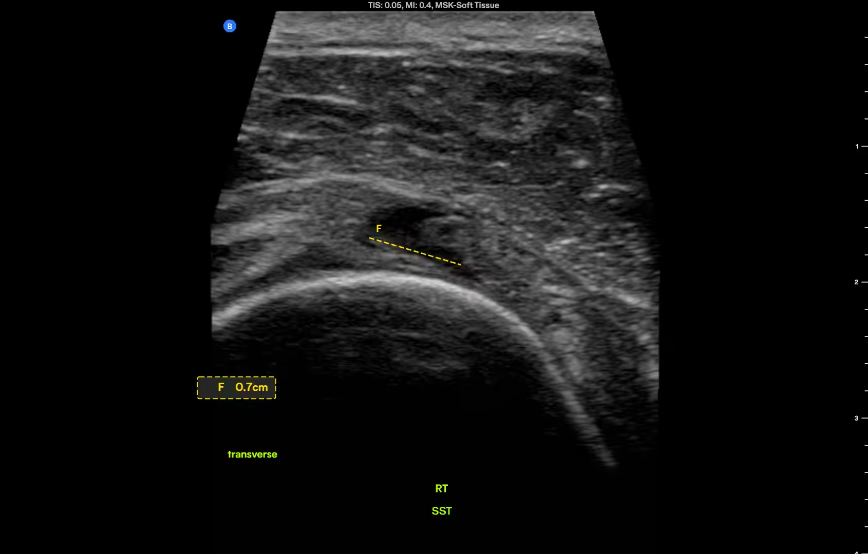
Long axis over the lateral shoulder showed the expected layers: skin, subcutaneous fat, deltoid with its parallel fiber pattern, the brighter supraspinatus tendon beneath it, and the hyperechoic humeral cortex at the base. An irregular anechoic cleft cut through the tendon near its humeral attachment, appearing nearly full thickness in that plane. Rotating the probe 90 degrees confirmed a full-thickness gap from the superficial to the deep edge, with a maximum tear width of 0.7 cm. Sweeping across the injury showed the defect running obliquely across more than half the tendon's roughly 2.5 cm width, with some posterior fibers still intact.
Outcome
Same-day referral to orthopedics and a quicker return to work. The pathway this patient avoided: weeks of rest and physical therapy that were never going to heal this, then more weeks negotiating MRI authorization. All the while the muscle atrophies and retracts, making repair technically harder.
Why the scan changed this case
A normal X-ray only meant no fracture. Ultrasound imaged the tendon in the office and diagnosed the high-grade tear. The grade sent this patient to a surgeon instead of physical therapy.
Why an ultrasound-first approach to MSK
“It’s immediately available, it’s safe, there’s no radiation, and it’s inexpensive relative to a CT or an MRI. The accuracy is really good too. I’ve become a real believer in taking that approach for a lot of the MSK problems I see in primary care. Being able to tell the patient right away and get him to orthopedics, rather than wasting six weeks of conservative care that isn’t going to work.”
Teaching point
Normal tendon is uniformly hyperechoic; loss of that echotexture is the finding. Two orthogonal planes are the minimum and neither is sufficient alone. Long axis suggested a near-full-thickness cleft, transverse confirmed it, and only sweeping the footprint revealed how far it ran.
Want to build this skill? Here’s where to start
Shoulder first, knee second. Not because other joints matter less, but because they are the highest-frequency presentations and the best place to build pattern recognition. Master tendinosis, joint effusion, tendon and ligament tears, and nerve entrapment first, then expand outward.

Explore the MSK case study series
Frequently asked questions
How is POCUS used in MSK specialty?

POCUS in MSK serves two purposes: diagnosing at the bedside and guiding procedures in real time. Diagnostically, iQ3 evaluates tendon and ligament injuries, joint effusions, nerve entrapments, muscle tears, and soft tissue masses — delivering answers in the room. On the procedural side, it guides joint injections and aspirations, bursa injections, nerve blocks, PRP delivery, and abscess drainage with real-time needle visualization.
iQ3 delivers both capabilities in one pocket-sized device — no cart, no probe swap, no separate imaging suite.
Which MSK presets does iQ3 offer, and how do they differ?
iQ3 includes 4 MSK-specific presets:
MSK – Soft Tissue delivers high dynamic range for soft tissue detail, covering 1–12 cm depth. It transitions automatically from linear to curvilinear format, spanning superficial trigger finger assessment to deep hip joint interrogation.
Musculoskeletal emphasizes contrast resolution for crisp structure and needle borders. Preferred for interventions and tendon or ligament evaluation.
Nerve is optimized for peripheral nerve assessment — nerve swelling, fascicular detail, and surrounding vessels — for carpal and cubital tunnel evaluation and hydrodissection guidance.
Needle: Out-Of-Plane
The Needle: Out-of-Plane preset is designed to help you see the needle tip sooner—within 1mm from the point of entry—when performing short-axis (out-of-plane) insertions.
Correct preset selection also reduces anisotropy artifacts, which can make normal tendons appear hypoechoic and mimic pathology.
Can Butterfly iQ3 guide MSK injections and procedures?
Yes. Procedural guidance is the fastest-growing MSK POCUS application, and iQ3 offers two features purpose-built for it.
NeedleViz™ colorizes the needle in real time at any approach angle, directly addressing the most common procedural frustration: maintaining tip visibility throughout the approach.
Biplane imaging shows simultaneous in-plane and out-of-plane views on one screen, confirming needle shaft and tip in both planes without repositioning — a first for handheld ultrasound.
Together, they reduce failed first-pass injections, lower cognitive load, and support safer needle delivery across joint injections, nerve blocks, PRP, and abscess drainage.
NeedleViz is a visualization aid and does not replace clinical judgment during ultrasound-guided procedures.
Can I use one probe for both MSK diagnosis and procedures?
Yes. iQ3 replaces three to four traditional transducers with a single whole-body probe. Its 1–12 MHz range covers superficial tendon and nerve imaging through deep joint and nerve block targets — no probe switch required.
In a typical encounter, a clinician can diagnose a rotator cuff abnormality and guide the injection — same probe, same device, same visit. The swappable USB-C/Lightning connector supports iOS and Android; the 8–10 hour battery covers a full clinic day.
From sidelines to clinics, the same device evaluates an acute injury and, when needed, supports aspiration or injection guidance.
What are the advantages of POCUS over MRI for MSK assessment?
POCUS and MRI are complementary tools. MRI remains the reference standard for comprehensive joint imaging and surgical planning. POCUS offers distinct bedside advantages:
Real-time and dynamic. Captures tendon movement, effusion dynamics, and nerve response during range of motion — none visible on MRI.
Immediate at the bedside. Provides answers in the room without scheduling or transport. POCUS often determines whether a finding warrants MRI at all.
Guidance capability. MRI cannot guide a needle; POCUS is the standard modality for real-time procedural guidance.
No contraindications related to implants or claustrophobia. Safe for patients with implants or claustrophobia.
For complete rotator cuff tears, POCUS can approach MRI accuracy in trained hands. Its practical role is to answer the immediate question — effusion, tendon integrity, nerve entrapment — and decide whether advanced imaging is needed.
Any specific accuracy comparisons to MRI to be reviewed and cleared by BN Regulatory.
Is Butterfly iQ3 useful for sports medicine and sideline assessment?
Yes. iQ3 and its MSK presets are built for sports medicine environments. At under 300g, it fits in a lab coat or equipment bag and moves from sideline to clinic without a cart.
On the sideline, iQ3 assesses tendon and ligament injuries, fractures, effusions, and muscle tears in real time, informing return-to-play or transport decisions. The same device then supports guided procedures when the patient follows up in the clinic.
Dynamic imaging under load reveals tendon and ligament function that static imaging cannot. Retrospective Cine Capture saves the video loop automatically, even without pressing record at the right moment.
How does Butterfly support MSK documentation, workflow, and billing?
Documentation and billing uncertainty are among the most-cited barriers to MSK POCUS adoption. Compass AI™ is designed to address both.
At the point of care: images and clips are captured, annotated, and saved to Butterfly Cloud automatically — eliminating the manual upload step that practitioners cite as a primary friction.
For billing: ultrasound-guided procedures require documented permanent image storage for CPT billing. Compass AI provides the structured archiving and reporting that makes this straightforward.
For practices and programs: Compass AI connects to the EHR and gives administrators visibility into scan volume, QA, and provider activity across sites — supporting credentialing and enterprise-wide adoption.
For clinicians: scan, document, and share without leaving the patient’s side. For practice owners: billable in-office procedures are captured and coded without the manual steps that get skipped in a busy clinic.
Compass AI™ is not intended to provide information used to make decisions regarding diagnosis or therapeutic purposes.
How long does it take to become proficient in MSK POCUS with iQ3?
MSK POCUS has a longer learning curve than cardiac or abdominal applications — dynamic scanning, anisotropy management, and simultaneous probe-and-needle control all add complexity. The curve is well documented and manageable with structured training.
Common applications — guided injections, aspirations, and basic tendon assessment — are typically achievable after a targeted CME course plus 25–50 supervised procedures per application.
Butterfly supports the learning process through: MSK-specific presets, in-app voice annotation, and a free introductory MSK course through Butterfly ScanLab education platform. Auto-optimization also reduces manual gain and depth adjustments for new users.
AI-assisted tools lower the expertise barrier, making MSK scanning accessible to trained NPs, PAs, and primary care physicians looking to expand their scope.
1. de Jesus JO, et al. AJR Am J Roentgenol. 2009;192(6):1701-1707.
2. Holtby R, Razmjou H. J Orthop Sports Phys Ther. 2004;34(4):194-200.